肾脏血管瘤
Haemangioma of the kidney
夏成青 赵明 程亮
发布时间:2016-04-03 06:54:31
概述:
发病部位:肾脏
诊断要点:
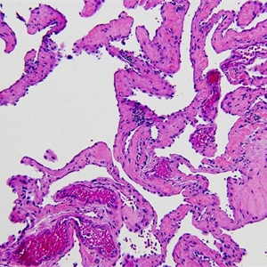
2.肾盏和肾盂最常受累,发生于肾皮质和肾被膜者较少见,肿瘤常较小;肿瘤无包膜,呈红色海绵状或红色条纹状;
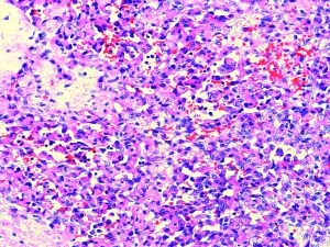
3.形态学包括毛细血管瘤和海绵状血管瘤;
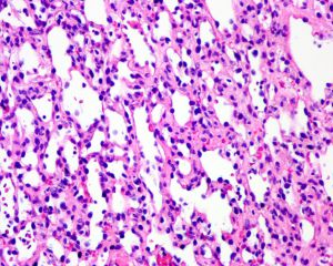
4.最常见为毛细血管瘤,表现为界限清楚的毛细血管型血管增生,可有肾小管陷入肿瘤中;
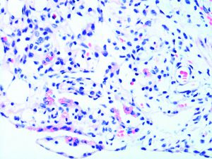
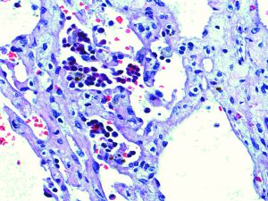
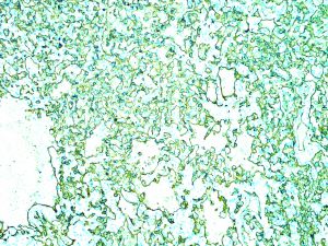
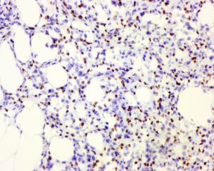
5.部分病例表现为交织状血管瘤(anastomosing hemangioma),常见发生于终末期肾疾病,血管腔相互吻合沟通,被覆鞋钉样瘤细胞,由少细胞的纤维性间质支撑,似脾窦结构,无明显的细胞异型性,罕见核分裂象。
6.可见血管内生长,血栓沉积,细胞内外的玻璃样小体以及髓外造血等。
免疫组织化学染色:
分子标记:
鉴别诊断:
伴有血管瘤样间质的低级别透明细胞肾细胞癌:瘤细胞稀少而间质血管丰富,类似于交织状血管瘤,免疫组化染色PAX8,CK,EMA可勾勒出血管性间质内少量的肿瘤细胞。
肾脏血管母细胞瘤:瘤细胞可见富于脂质的多泡状空泡状胞浆,间质血管网丰富,免疫组化染色肿瘤细胞表达Inhibin,NSE和S100蛋白,不表达血管内皮标志物。
血管肉瘤:罕见发生于肾脏,呈侵袭性的浸润性生长,至少局灶可见明显的瘤细胞异型性和核分裂象包括非典型核分裂象。
预后:
治疗:
参考文献:
Montgomery E, Epstein J I. Anastomosing hemangioma of the genitourinary tract: a lesion mimicking angiosarcoma [J]. Am J Surg Pathol, 2009, 33(9): 1364-9.
Anastomosing hemangioma of the genitourinary system: eight cases in the kidney and ovary with immunohistochemical and ultrastructural analysis [J]. Am J Clin Pathol, 2011, 136(3): 450-7.
Kryvenko O N, Roquero L, Gupta N S, et al. Low-grade clear cell renal cell carcinoma mimicking hemangioma of the kidney: a series of 4 cases [J]. Arch Pathol Lab Med, 2013, 137(2): 251-4.
Kryvenko O N, Haley S L, Smith S C, et al. Haemangiomas in kidneys with end-stage renal disease: a novel clinicopathological association [J]. Histopathology, 2014, 65(3): 309-18.
赵明,孔梅,余晶晶,何向蕾,张大宏,滕晓东. 肾脏及肾上腺交织状血管瘤临床病理分析. 中华病理学杂志. 2016;45(10):698-702.
