血管脂肪瘤
Angiolipoma
刘正智 赵明
发布时间:2016-07-27 13:10:26
概述:
发病部位:前臂,躯干和上臂。
诊断要点:
1. 常见于青年人,好发于前臂、躯干和上臂皮下,有包膜;
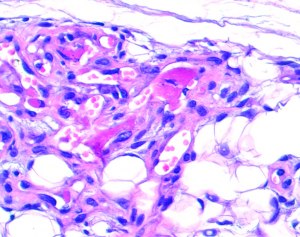
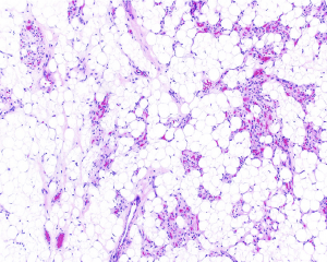
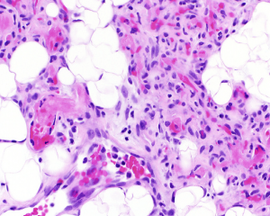
2. 镜下可见肿瘤主要由脂肪和分支状的毛细血管网组成,两者比例因人而不同;
3. 包膜下区域血管密度较高,小血管内常见纤维素性微血栓;
4. 脂肪分化成熟,与血管交叉分布,间质内可见肥大细胞浸润;
5. 少数病例以血管为主(>90%),并伴有较多血管周的梭形细胞(肌周皮细胞),而脂肪成分相对稀少,称为富于细胞性血管脂肪瘤。
免疫组织化学染色:
分子标记:
鉴别诊断:
2、Kaposi肉瘤:更多的梭形和束状内皮细胞增生;充血狭缝样空间和渗出性红细胞 ;PAS(+)透明球,但无纤维蛋白血栓 ;HHV8(+)内皮细胞。
3、梭形细胞血管瘤:好发于肢端多发性皮下结节;与细胞血管脂肪瘤不同,SCH有大、海绵状血管空间 ;纺锤状和上皮样内皮细胞的细胞区和类似微脂肪细胞的聚集内皮泡;大钙化血栓(静脉),但无聚集血管,血栓小。
治疗:
参考文献:
1. Sheng W et al: Cellular angiolipoma: a clinicopathological and immunohistochemical study of 12 cases. Am J Dermatopathol. 35(2):220-5, 2013
2. Bang M et al: Ultrasonographic analysis of subcutaneous angiolipoma. Skeletal Radiol. 41(9):1055-9, 2012
3. Ghosal N et al: Angiolipoma in sellar, suprasellar and parasellar region: report on two new cases and review of literature. Clin Neuropathol. 30(3):118-21, 2011
5. Arenaz Búa J et al: Angiolipoma in head and neck: report of two cases and review of the literature. Int J Oral Maxillofac Surg. 39(6):610-5, 2010
6. Dimosthenous K et al: Angiolipoma of the thyroid gland. Int J Surg Pathol. 17(1):65-7, 2009
7. Lapidoth M et al: Capillary malformation associated with angiolipoma: analysis of 127 consecutive clinic patients. Am J Clin Dermatol. 9(6):389-92, 2008


