软组织神经束膜瘤
Soft Tissue Perineurioma
赵明 刘正智
发布时间:2016-08-02 14:39:00
概述:
发病部位:好发于四肢和躯干的浅表软组织。
诊断要点:
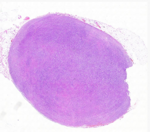
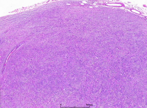
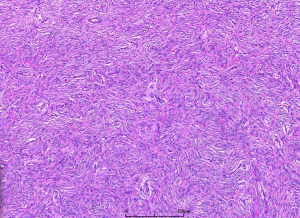
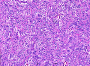
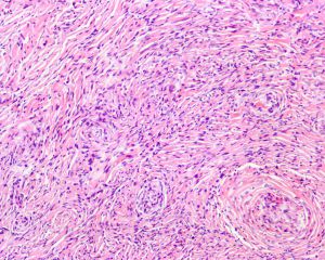
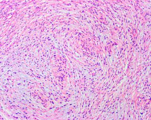
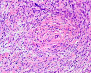
2. 镜下见纤细的纤维母细胞样梭形细胞呈束状或波浪状排列,有时呈疏松的漩涡状或模糊的席纹状排列;瘤细胞无异型,罕见核分裂象;
3. 部分病例富于细胞,瘤细胞多呈明显的席纹状排列,间质较少;
4. 部分病例细胞稀疏,呈星状向黏液性间质内伸出细长的突起,间质含有大量的黏液;
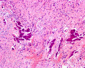
5. 少数病例间质显著胶原化,瘤细胞呈纤细的梭形,并为粗大的胶原纤维所分隔,偶见钙化和砂粒体形成;
6.硬化性神经束膜瘤男性居多,好发于手指;大多数类型男女比例相等;
7.本瘤有高达30%的病例可出现在深部组织或内脏。
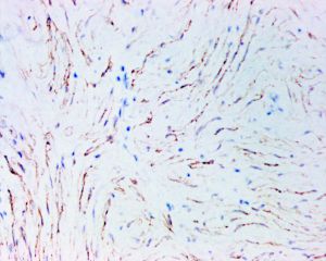
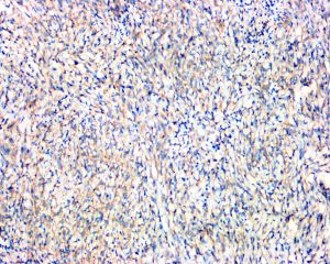
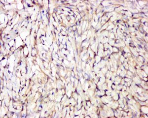
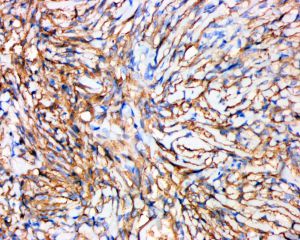
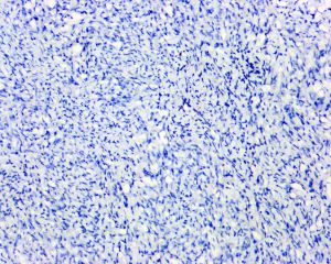
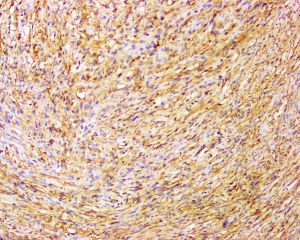
免疫组织化学染色:
分子标记:
鉴别诊断:
1、神经纤维瘤:S100(+)。
2、神经鞘瘤:有明显的Antoni A and B区,S100(+),EMA、GLUT-1(-)。
3、隆突性皮肤纤维肉瘤:瘤细胞向皮下浸润性生长,CD34(+),EMA(-), claudin-1(-),特征性Tt(17;22)异位涉及 COL1A1 and PDGFB。
4、孤立性纤维性肿瘤:瘤细胞漩涡生长,无固定方向,特征性的“鹿角”状血管;CD34、STAT6(+),MEA、GLUT-1(-)。
治疗:
参考文献:
1.Agaimy A et al: Comparative study of soft tissue perineurioma and meningioma using a five-marker immunohistochemical panel. Histopathology. 65(1):60-70, 2014
2.Santos-Briz A et al: Cutaneous intraneural perineurioma: a case report. Am J Dermatopathol. 35(3):e45-8, 2013
3.Agaimy A et al: Benign serrated colorectal fibroblastic polyps/intramucosal perineuriomas are true mixed epithelial-stromal polyps (hybrid hyperplastic polyp/mucosal perineurioma) with frequent BRAF mutations. Am J Surg Pathol. 34(11):1663-71, 2010
4.Ferraresi S et al: Perineurioma of the sciatic nerve: a possible cause of idiopathic foot drop in children: report of 4 cases. J Neurosurg Pediatr. 6(5):506-10, 2010
5.Fox MD et al: Extra-acral cutaneous/soft tissue sclerosing perineurioma: an under-recognized entity in the differential of CD34-positive cutaneous neoplasms. J Cutan Pathol. 37(10):1053-6, 2010
6.Groisman G et al: Early colonic perineurioma: a report of 11 cases. Int J Surg Pathol. 18(4):292-7, 2010
7.Koutlas IG et al: Intraoral perineurioma, soft tissue type: report of five cases, including 3 intraosseous examples, and review of the literature. Head Neck Pathol. 4(2):113-20, 2010
8.Hornick JL et al: Hybrid schwannoma/perineurioma: clinicopathologic analysis of 42 distinctive benign nerve sheath tumors. Am J Surg Pathol. 33(10):1554-61, 2009
